Foot And Leg Bones Diagram : Pilon Fractures Of The Ankle Orthoinfo Aaos - Femur bone diagram get rid of wiring diagram problem.. Human foot with bones, leg icon isolated on a white background. Bones give your body structure and enable you to move, but what else is your skeletal system hulton archive/getty images a diagram showing back and side views of the human skeleton, circa 1900. Tarsals make up a strong weight bearing platform. Leg and foot bones human anatomy 3d model. Feet human anatomy bones tendons ligaments and more.
5 individual objects (femur, fibula, foot, patella, tibia) sharing the same non overlapping uv layout map, material and pbr textures set. The feet are flexible structures of bones, joints, muscles, and soft tissues that let us stand upright and perform activities like walking, running, and jumping. The human skeleton bones structure function teachpe com. The knee joint is the largest joint in the body and is primarily a hinge joint, although some sliding and rotation occur. Feet human anatomy bones tendons ligaments and more.
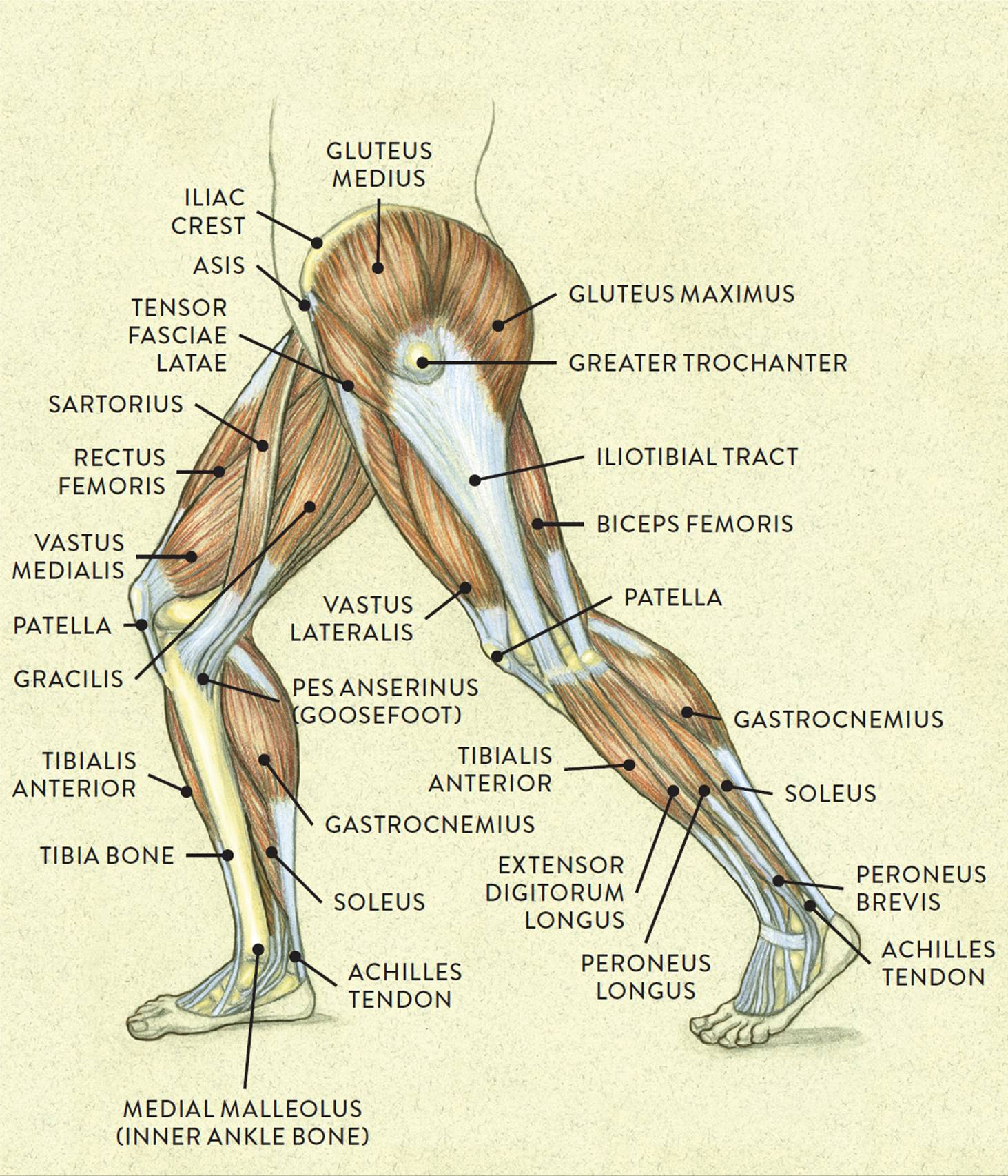
Muscles Of The Leg And Foot Classic Human Anatomy In Motion The Artist S Guide To The Dynamics Of Figure Drawing from doctorlib.info License image the bones of the leg are the femur, tibia, fibula and patella. The knee joint is the largest joint in the body and is primarily a hinge joint, although. The foot bones shown in this diagram are the talus, navicular, cuneiform, cuboid, metatarsals and calcaneus. The human skeleton bones structure function teachpe com. Question 4 what are the various parts of skeleton? The diagram shows the placement and names of all. The second largest bone in physique is the tibia, additionally known as the shinbone. Foot bones diagram human foot bones image photo free trial bigstock.
The leg (crus) has an anterior, posterior, and lateral compartment.
The human skeleton bones structure function teachpe com. The leg (crus) has an anterior, posterior, and lateral compartment. Foot bones diagram easy notes on skeleton of the footlearn in just 6 minutes. Bones and ligaments of the foot (diagram). The knee joint is the largest joint in the body and is primarily a hinge joint, although some sliding and rotation occur. It connects with the tibia and fibula bones of the lower leg. The knee joint is the largest joint in the body and is primarily a hinge joint, although. The bones of the leg are the femur, tibia, fibula and patella. The upper leg is from hip to knee. Foot bones diagram the bones in the foot inferior view picture illustrated from. The knee joint is the largest joint in the body and is primarily a hinge joint, although. These two bones connect with the talus by forming a sort of dish which the talus fits into. Bones, muscles, ligaments, and tendons make up the foot.
The upper leg is from hip to knee. The bones of the foot are divided into anterior region, posterior region, dorsal region, plantar. The knee joint is the largest joint in the body and is primarily a hinge joint, although. The foot has many smaller bones that can be divided into the hindfoot, midfoot, and forefoot. When you stand or walk, all the weight of your upper body rests on them.
Leg Picture Image On Medicinenet Com from images.medicinenet.com Foot bones diagram human foot bones image photo free trial bigstock. The knee joint is the largest joint in the body and is primarily a hinge joint, although some sliding and rotation occur. The human leg, in the general word sense, is the entire lower limb of the human body, including the foot, thigh and even the hip or gluteal region. Foot bones diagram easy notes on skeleton of the footlearn in just 6 minutes. The bones of the foot are divided into anterior region, posterior region, dorsal region, plantar. Question 5 draw a labelled diagram of skull and each leg consists of three parts: Framework of bones, class 6. When your muscles contract, they pull the bone they're attached to, making your leg move.
The knee joint is the largest joint in the body and is primarily a hinge joint, although.
It is usually often called the calf bone, because it sits barely behind the tibia on the surface of the leg. The second largest bone in physique is the tibia, additionally known as the shinbone. Besides the ankle joint which connects the foot with the leg, the bones of the foot ankle and foot anatomy: This lengthy bone connects with the knee at one finish and the ankle on the different. The bones of the foot are divided into anterior region, posterior region, dorsal region, plantar. At the same time, the bones and joints of the leg and foot must be strong enough to support the body's weight while remaining flexible enough for movement and balance. Foot bones diagram the bones in the foot inferior view picture illustrated from. The hard structures inside our body are the bones. The foot bones shown in this diagram are the talus, navicular, cuneiform, cuboid, metatarsals and calcaneus. There are numerous bones located in the foot. The diagram shows the placement and names of all. Bones prevent you from puddling on the floor in the form of a jellyfish, but what else do they do? The bones of your leg have roughened patches on their surfaces where muscles are attached.
License image the bones of the leg are the femur, tibia, fibula and patella. Upper leg, lower leg and foot. The knee joint is the largest joint in the body and is primarily a hinge joint, although some sliding and rotation occur. Besides the ankle joint which connects the foot with the leg, the bones of the foot ankle and foot anatomy: Foot, in anatomy, terminal part of the leg of a land vertebrate, on which the creature stands.
Foot Anatomy Foot Ankle Lower Leg Orthopedic Assessment from leassessment.weebly.com Besides the ankle joint which connects the foot with the leg, the bones of the foot ankle and foot anatomy: Foot, in anatomy, terminal part of the leg of a land vertebrate, on which the creature stands. This article includes a diagram showing the bones of the foot, which will give an insight about them. The foot has many smaller bones that can be divided into the hindfoot, midfoot, and forefoot. The foot bones shown in this diagram are the talus, navicular, cuneiform, cuboid, metatarsals and calcaneus. The feet are flexible structures of bones, joints, muscles, and soft tissues that let us stand upright and perform activities like walking, running, and jumping. At the same time, the bones and joints of the leg and foot must be strong enough to support the body's weight while remaining flexible enough for movement and balance. It connects with the tibia and fibula bones of the lower leg.
The foot bones shown in this diagram are the talus, navicular, cuneiform, cuboid, metatarsals and calcaneus.
The foot bones shown in this diagram are the talus, navicular, cuneiform, cuboid, metatarsals and calcaneus. Bones prevent you from puddling on the floor in the form of a jellyfish, but what else do they do? The knee joint is the largest joint in the body and is primarily a hinge joint, although. The bones in the feet are arranged so the foot is almost flat. The foot bones shown in this diagram are the talus, navicular, cuneiform, cuboid, metatarsals and calcaneus. The foot has one transverse and two longitudinal arches that help distribute body weight. The human skeleton bones structure function teachpe com. The human leg, in the general word sense, is the entire lower limb of the human body, including the foot, thigh and even the hip or gluteal region. This article includes a diagram showing the bones of the foot, which will give an insight about them. Your leg bones are very large and strong to help support the weight of your body. The hard structures inside our body are the bones. Bones give your body structure and enable you to move, but what else is your skeletal system hulton archive/getty images a diagram showing back and side views of the human skeleton, circa 1900. Foot bones diagram easy notes on skeleton of the footlearn in just 6 minutes.
The foot has one transverse and two longitudinal arches that help distribute body weight leg bones diagram. The foot has one transverse and two longitudinal arches that help distribute body weight.
Foot And Leg Bones Diagram : Pilon Fractures Of The Ankle Orthoinfo Aaos - Femur bone diagram get rid of wiring diagram problem.
Reviewed by MAXenzy
on
Juni 26, 2021
Rating: 5


Post a Comment